Photographs
Geitler used photographs sparingly, but his classic 1932 work on the diatom life cycle includes photographs illustrating the frustules and colonies of Eunotia, pairing and auxospore formation in Cocconeis, the curious incunabula present around the auxospore in Nitzschia fonticola, the perizonium of Epithemia, etc. Anyone who has attempted to use photography to record sexual reproduction and auxosporulation in diatoms will know how difficult it is to capture the essential features of each stage in one or a few shots. If the nuclei are in focus, then probably the chloroplasts are not: the depth of focus of conventional light microscopes is small, compared to the size of diatom cells. Hence it is not surprising that Geitler usually preferred to illustrate his findings with drawings, and these are always models of clarity, combining information economically from many different focal planes.
Close examination of some of the original photographs for the
1932
work suggests that, although Geitler thought the photographs were worth
including, he was sometimes frustrated by being unable to produce
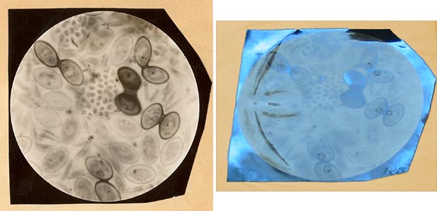
images that combine clarity with complexity. For example the image on
the left above, which was reproduced as Figure 75 in the published
monograph, shows many Cocconeis
cells, some of them vegetative (the paler, single cells) and some of
them sexual (there are five copulating pairs). It would have been
tedious to draw such a complex image and it was obviously better to use
a photograph. However, Geitler also wished to show cytological detail
in the paired cells, to show how the nuclei were displaced towards each
other. But although he could see this in his microscope, the
photograph did not show it adequately. Geitler's solution was one that
we might now regard as improper: he drew in ink on the photograph,
reinforcing the outlines of the nuclei and adding density to the
nucleoli. This can be seen in the right-hand image above, which is an
oblique view of the same photograph shown on the left. Similar
treatment
was given to Figure 119, reinforcing the irregular transverse banding
of
the incunabula in Nitzschia
fonticola, which he also drew in Figure 121.
In neither case was anything added that could not be seen in the
original material, but the published images are nevertheless deceptive.
Reference: L. Geitler (1932). Der Formwechsel der pennaten Diatomeen. Archiv für Protistenkunde 78: 1–226.
The caption to Figure 75 in Geitler (1932) states: "... Detailbild von fünf Copulationspaaren: vier vor, das mittlere während der Copulation. – Mikrophoto, Zeiss Apo 90, 1.30, Reichert-Aufsatzkamera."
David Mann, August 2009


