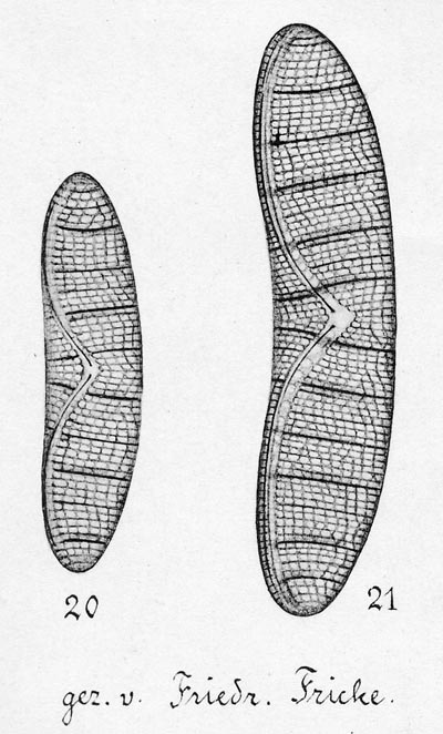
Epithemia valves drawn by Friedrich Fricke in a later part of Adolf Schmidt's Atlas der Diatomaceenkunde, published in 1904. Epithemia valves, like Petroneis valves, have transverse lines of pores. However the pores are larger and more complex than in Petroneis and it is very rare for them to be drawn. Instead, the ribs separating the pores are shown. The same choice has to be made in photomicroscopy: should the image appear to portray the pores, or the ribs and frets that surround and create the pores?