Introduction to diatoms: discovery
Discovery
Diatoms are generally microscopic organisms. The largest, by volume, are the giant planktonic diatoms Ethmodiscus gazellae and E. rex, which occur in the plankton of warm oceans. These can be up to 2 mm in diameter and are easily visible to the naked eye as small cylinders, about as high as wide, but most diatoms are much smaller – less than 100 µm in their maximum dimension. Not surprisingly, therefore, diatoms were unknown before the invention of microscopes.
In the late 17th century several European scientists began to use early simple or compound microscopes to look at natural material from aquatic habitats. Among them was Anthonie van Leeuwenhoek, who used single bead-like lenses and discovered protists ('animalcules'), sperm and bacteria. Leeuwenhoek almost certainly saw diatoms – they are certainly present in some of the samples he took (Ford 1991) – but none of his published illustrations can be convincingly argued to show diatoms.
In 1703, another early microscopist sent a paper to the Royal Society of London, which was published in its Philosophical Transactions. His name was not recorded, either on the paper itself or in the Royal Society archives, and the paper was communicated to the Royal Society via another unnamed person referred to as 'Mr. C', who could be any one of four Fellows. For many years the authorship of the 1703 paper (and of another by the same 'Gentleman') remained a mystery but in 2019 historical research by John Dolan revealed his identity – Charles King, a hitherto unrecognized microscopist and proto-protistologist born in 1654.
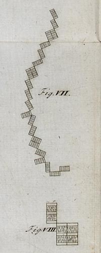
There was never much mystery, though, about what the 'anonymous' microscopist saw: King's drawing is a careful and lovely representation of the freshwater diatom Tabellaria, shown left. Tabellaria forms colonies of cells connected by their corners. Benthic Tabellaria, growing attached to plants or rocks usually form extended zig-zag chains (with some 'fans' of three cells, as in two cases here), whereas planktonic species form star-shaped colonies. Each of the rectangles here is a single cell, while the squares are pairs of cells, produced by division; later, the daughter cells will split apart, remaining attached by a 'hinge' of polysaccharide at one end. The three white 'stripes' on each rectangle are created by the shape of the cell (in the plane at right-angles to the view shown), which is expanded at the centre and ends.
The image is © The Royal Society
References
Anonymous (1703). Two letters from a gentleman in the country, relating to Mr. Leuwenhoeck’s letter in Transaction, No. 283. Philosophical Transactions of the Royal Society of London 23 (288): 1494
Dolan, J. (2019). Unmasking "The Eldest Son of The Father of Protozoology": Charles King". Protist 170: 374-384.
Ford, B.J. (1991). The Leeuwenhoek legacy. Biopress and Farrand Press, 185 pp.