Background
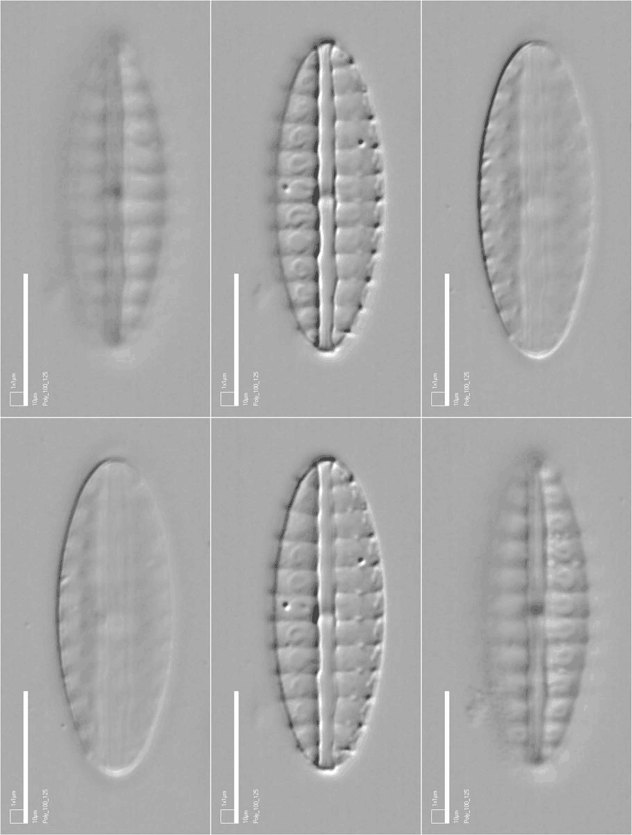
In a publication it is usually only possible to illustrate a specimen with one or sometimes a few photographs. Because of the very limited depth of focus of high-magnification optical microscopy, a single photograph is usually not enough to show all the detail that can be seen by focusing up and down through a specimen.
However, using the WWW, whole specimens can be viewed as stacks of optical sections that your browser opens sequentially. What you see is rather like the Diploneis specimen on the right, except that most of the specimens do not loop automatically. Instead, we supply a focusing control so that you can look through the whole specimen interactively, or we provide a video file.
Photographs
The photographs of Sellaphora and Diploneis types were taken on a Reichert Polyvar 2 compound microscope using a Polaroid DMC2 digital camera, with a resolution of 1600 x 1200 pixels. Individual optical sections of the Sellaphora specimens were taken at a vertical interval of 0.2 Ám as measured by the calibrated fine-focus control of the Polyvar, but since the refractive index of the naphrax in which the specimens were mounted is higher than that of glass, this corresponds to more than 0.2 Ám of the specimen.
The Polyvar fine-focus control is calibrated in steps of 1 Ám. To allow us to take photographs at closer intervals, we made and attached a vernier scale to the fine focus.
A scale bar was added to each photograph using the image-analysis program Optimas. The scale bar includes a 1 x 1 Ám square to enable one to check whether or not the pixel aspect ratio has been changed at any stage in the display process (if the 'square' is not square, then the pixel aspect ratio is wrong). Brightness, contrast and image size were all adjusted using Adobe PhotoShop.
More recently, we have acquired a Zeiss Axio-Imager microscope with
a computer-controlled motorized z-control and camera. With this we can
acquire image stacks much more rapidly than with the Polyvar, at much
smaller vertical intervals.
Web pages and focus bar
The focus bar was created using a JavaScript program available on the web (thanks are due to Christiaan Hofman for producing the code, and to Paul Rosen for directing us to it). The bar may not work using some browsers and so we also present the image stacks as .avi video files [using VideoMach software (available from www.gromada.com)]. These are best viewed using QuickTime, because the progress button can be grabbed and moved by mouse, just like the JavaScript slider. Some other players are less versatile.