Amphora copulata: diakinesis
- image stack with slider bar for focus. Note the particularly long bivalent at left-centre.
- gif image
- avi video file [we strongly recommend that you use QuickTime for this, because it will allow you to select individual frames or move backwards or forwards through the sequence]
- images of one gametangium and the other gametangium (focused on the valves; scale bars = 10 µm)
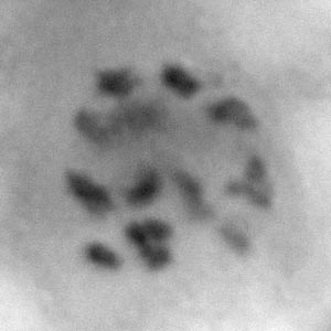
Amphora ovalis: diakinesis
- image stack with slider bar for focus. This shows a single diakinetic nucleus; our estimate is 14 or 15 bivalents.
- avi video file (CAUTION! 4 Mb) In contrast to the stack in the slider bar version, the video clip shows both gametangia, which were at the same stage of meiosis. [we strongly recommend that you use QuickTime].
Amphora ovalis: diakinesis/prometaphase I
- image stack with slider bar for focus. Metacentric and acro- or telocentric chromosome bivalents are visible
- gif image
- avi video file [we strongly recommend that you use QuickTime]
Amphora ovalis: diplotene/diakinesis
- image stack with slider bar for focus. Metacentric and acro- or telocentric chromosome bivalents are visible
- gif image (CAUTION! 3 Mb)
- avi video file [we strongly recommend that you use QuickTime]
Amphora minutissima: diakinesis and following meiosis II (showing 4-nucleate stage with 2 nuclei degenerating)
The upper gametangium is in early diakinesis: the chromosomes have not separated as widely as in the A. copulata example. In the lower gametangium, which has recently completed telophase of meiosis II, four nuclei are visible. Two are compact and very densely staining, and have already begun to degenerate: they will remain visible as steadily shrinking but dark-staining 'pyknotic' nuclei until well after auxospore expansion has begun. The other two nuclei are more diffuse and are regaining the interphase condition of dispersed chromatin and heterochromatic granules.
- image stack with slider bar for focus
- gif image (CAUTION! 3.5 Mb)
- avi video file [we strongly recommend that you use QuickTime]