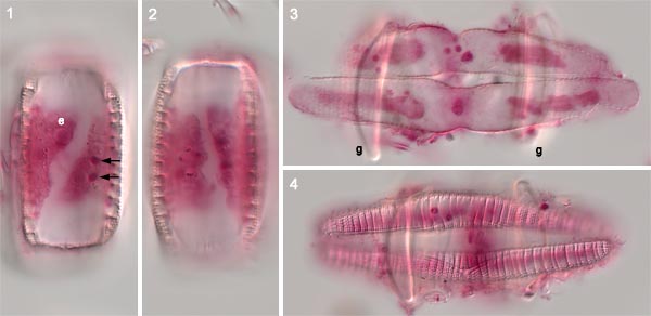
These photographs were taken in 2013 of cells fixed (with formalin) during the German ‘Sunda’ expedition of 1928–9 and subsequently stained with carmine before 1932 by Lothar Geitler. The preparation used was Sunda sample R1c from Sumatra (Geitler 1932, p. 158; 1977). The photographs were taken in 2013, showing the high permanence of carmine staining (see also Mann 2015).
The gametangia and auxospores contain endosymbiotic cyanobacteria (e.g. e). The two gametangia shown in Figs 1 and 2 exhibit a curious feature, thought to be characteristic of Epithemia and Rhopalodia: after meiosis II, the two gametic protoplasts become partly rearranged within the gametangia so that the plane separating them is diagonal within the cell. In other pennate diatoms rearrangement either does not occur at all, or is completed, so that the gametes lie one towards each end of the cell.
The auxospores show another feature that is unusual in pennate diatoms: they are constricted at the centre (Fig. 3).
Auxospore expansion takes place at right angles to the gametangia
(the gametangial valves [g] can be seen out of focus in Figs 3 and 4).
Each auxospore contains two chloroplasts, one derived from each gamete
(Fig. 3).


 This site is
hosted
by
the Royal Botanic
Garden Edinburgh.