Type specimens: images and data

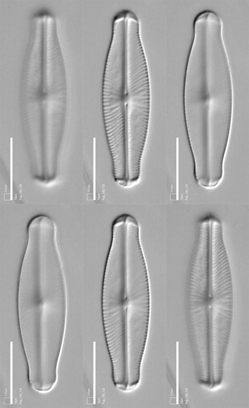
Image: type specimen of Sellaphora
lanceolata.
Types are used to determine
how the names of diatom species are to
be used, just as in other groups of organisms. Types are sometimes
individual specimens, mounted in resin on a microscope slide, but more
often they are whole slides, prepared from a mixed natural
population containing several or many specimens of the species that the
slide typifies, together with specimens of many other species. The use
of whole slides as types is allowed by the International Code of
Botanical Nomenclature, but it would be much better to designate a
single
specimen, to provide an unambiguous reference point for comparisons.
Some diatoms are typified by images.
Nowadays, when a species is described for the first time, the
author(s) of the description must specify a type specimen and this is
called the holotype. In the
past, this was not mandatory and so for species originally described
without a type, taxonomists have to choose a specimen (or slide) on
behalf of the original author. This may be a lectotype, if appropriate
specimens studied by the original author still survive, or a neotype,
if they do not. In addition, if the holotype, lectotype or neotype do
not exhibit enough characterictics to define the species adequately and
unambiguously, an epitype can be added. These procedures aim to
stabilize and clarify the use of the names that all other biologists
use for communication: though they appear arcane, they are important.
In AlgaeWorld, you can find information and images for some type
specimens. We have
produced stacks of images, by focusing through specimens manually or
with
a computerized microscope, and we present these stacks here so that you
can look at type specimens almost
as though
you were looking
down a microscope. By sliding a focus button or running a short video
sequence, you can focus up and down
through the specimens to see details that may be of importance
for identification and comparison –
in this way, we can give you much more than can be seen in a single
photograph.
Not all of the species that
we have described are illustrated here (principally those in the papers
by Droop
1998 and Mann
et al.
2004). Follow this link for technical
information.
- Cymbellonitzschia
minima
Hustedt
- Diploneis
sejuncta
(Schmidt) Jřrgensen (= Navicula
sejuncta
Schmidt)
- Diploneis
praesejuncta
S.J.M. Droop, sp. nov.
- Diploneis
solea
S.J.M. Droop, sp. nov.
- Diploneis
pneumatica
S.J.M. Droop, sp. nov.
- Diploneis
biremiformis
S.J.M. Droop, sp. nov.
- Diploneis
colae
S.J.M. Droop, sp. nov.
- Eunophora
berggrenii
(Cleve) D.G. Mann, M.A. Harper & Levkov
- Hantzschia segmentalis
Brun
- Lyrella cassiteridum D.G. Mann,
sp. nov.
- Navicula
pupula
Kützing
- Nitzschia abbreviata Simonsen
- Nitzschia brachygramma D.G.
Mann & Trobajo, sp. nov.
- Nitzschia dicrogramma D.G. Mann
& Trobajo, sp. nov.
- Nitzschia parkii D.G. Mann
& Trobajo, sp. nov.
- Sellaphora
auldreekie
D.G. Mann & S.M.
McDonald, sp. nov.
- Sellaphora
blackfordensis
D.G. Mann & S.
Droop, sp. nov.
- Sellaphora
capitata
D.G. Mann & S.M.
McDonald, sp. nov.
- Sellaphora
lanceolata
D.G. Mann & S.
Droop, sp. nov.
- Sellaphora
obesa
D.G. Mann & M.M. Bayer, sp. nov.
- Sellaphora
pupula
(Kützing) Mereschkowsky
David Mann
Royal Botanic Garden Edinburgh
September 2010, amended February 2014


 This site is hosted
by
the Royal Botanic
Garden Edinburgh.