The preparation from which these photographs were taken was prepared
in the 1980s (for the work reported by Mann 1988). The photographs were
taken in 2013, showing the high permanence of carmine staining (see also Mann 2015).
Parlibellus protracta
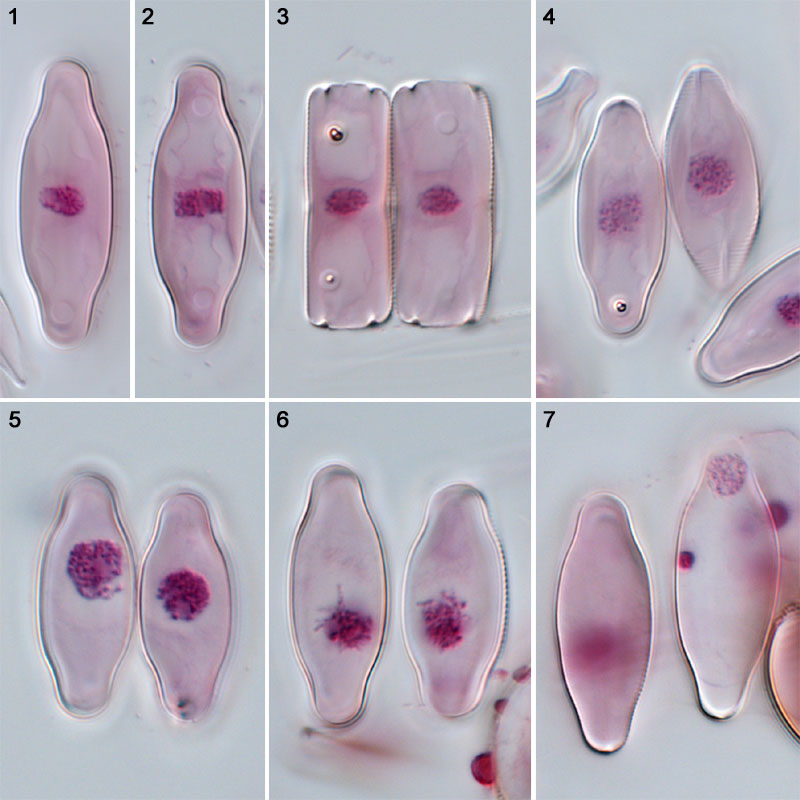
Figs 1–7. Parlibellus protracta:
acetocarmine stained, interference contrast optics.
Figs 1, 2. Vegetative cells in interphase, valve view.
Fig. 3. A recently divided vegetative cell, in girdle view.
Fig. 4. Paired cells: leptotene of meiotic prophase.
Figs 5, 6. Zygotene.
Fig. 7. Gametangia after meiosis II: each gamete contains a single functional haploid nuclues (at top in righthand cell) and a degenerating, densely stained nucleus (below).
Clicking on the image will load a enlarged version.
References:
Mann, D.G. (1988). Sexual reproduction and systematics of Navicula protracta. Diatom Research, 3: 227–236.
Mann, D.G. (2015). Unconventional diatom collections. Nova Hedwigia, Beiheft 144: 35–59.
David Mann,
Royal Botanic Garden Edinburgh
July 2013