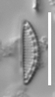
differential interference contrast optics
- pop-up with slider bar
- .avi movie (best viewed with QuickTime)

Scale bar = 10 µm
differential interference contrast optics
Ringed specimen on Hustedt slide 224/75 (Alfred Wegener Institut,
Bremerhaven). This frustule was shown to be the
type of Cymbellonitzschia minima
by R. Simonsen (1987) and was reexamined during work by Trobajo et al. (submitted).
There are several frustules of Cymbellonitzschia
minima
on the type slide, including some that are hantzschioid while others
are nitzschioid. Frustules lying in girdle view can be quite easily
confused with frustules of Nitzschia
epiphyticoides,
also present on slide 224/75, also exhibiting hantzschioid and
nitzschioid symmetries, and also with low striation densities (c. 20-24
in 10 µm). However, the two species can be distinguished in girdle view
by careful focusing, which reveals the dissimilar (strongly convex and
flat) sides of the frustule in Cymbellonitzschia,
but similar and only slightly convex sides in N. epiphyticoides. Here we
illustrate a nitzschioid frustule of C.
minima.
Trobajo, R., Mann, D.G. & Cox, E.J. (submitted). Studies on the type material of Nitzschia abbreviata (Bacillariophyta).
Simonsen, R. (1987). Atlas and catalogue of the diatom types of
Friedrich Hustedt, 3 vols. J. Cramer, Berlin & Stuttgart.
David Mann & Rosa Trobajo
Royal Botanic Garden Edinburgh
December 2010