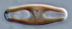
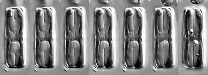
Sellaphora cell in girdle view, during cytokinesis
Diatom cells divide through the inward development of a cleavage furrow. In most freshwater diatoms, the cleavage furrow is extremely narrow and, when seen in mid-focus from the side (i.e. when the cells are in 'girdle view'), it appears as a pair of lines that grow in towards the centre, as seen here. The furrow appears at the poles and deepens, first cutting the vacuoles in half, and then bisecting the cytoplasmic bridge in which the mitotic nucleus lies. Finally (last photograph), the two daughter protoplasts are completely separated and lie side-by-side within the parent cell wall. At this stage, the daughter protoplasts are naked. Very soon, however, new valves begin to be formed within them (in special, membrane-bound sacs called 'silica deposition vesicles'). Once the valves are complete, they are secreted (exocytosed) from the daughter cells, which thereby acquire a functional cell wall. The daughter cells then separate.
In the last frame, note that the two volutin granules (the strongly refractile spheres) have been segregated one to each daughter cell.
On the next page, we show the same process, but rotated by 90º, i.e. in valve view.
The sequence shown spanned 4 minutes.


 This site is hosted by the Royal Botanic
Garden Edinburgh.