Transformation into auxospores
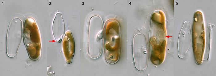
After the male gamete has fused with the female gamete and moved bodily through the copulation aperture, configurations are produced looking like Figs 1–3 above, with the zygote apparently filling the whole of the female gametangium. In fact, this isn't quite true, because the female gametangium also contains another cell – namely, the supernumerary cell produced in the gametangium by the unequal cytokinesis after meiosis I. Zygotes are always elongate and take the shape of the female gametangium – lanceolate or elliptical in such species as S. auldreekie (Fig. 2), linear in S. laevissima (Fig. 3), etc. The contents of the zygote are very densely packed and the female gametangium is visibly distended. Vacuoles are not obvious, and the two chloroplasts inherited from the gametes fold around each other and cannot be distinguished separately using normal LM optics. Two volutin granules are present in the zygote (visible in Fig. 3), and two are extruded and left behind by the male gamete in a small residual body at one end (Fig/ 2, arrow) or one side (Fig. 3) of the male gametangium. Why they are left behind is unknown.
The zygote is surrounded by a thin organic wall, but it is not a dormant stage. It remains the same size for several hours, during which its contents become rearranged and the nuclei become closely associated on one side at the centre (see 'Sexual reproduction IV', fig. 4), and then it begins to expand. The thecae of the female gametangium split apart and the zygote pushes out at each end (Figs 4 and 5). It is now considered to be an 'auxospore'. There is no change in ploidy during the transition fom zygote to auxospore; this is simply a cellular differentiation.
As the auxospore expands, it begins to generate the shape of the new generation of vegetative cells. In species that have tapering valves, the auxospore soon begins to taper (Fig. 5); in species with linear valves, the auxospore tends to maintain the same diameter; and in species with a central bulge, the diameter of the expanding tip first contracts and then stays ± constant (Fig. 4). During expansion, the chloroplasts spread out (when it can be seen that there are two), and the two gametic nuclei become more easily seen, lying in a pocket of cytoplasm at one side surrounded by the expanding vacuoles.It seems that it is the swelling of the vacuoles that drives auxospore expansion; in other diatoms it has been shown that transference to media of higher osmotic potential can halt expansion.


 This site is hosted by the Royal Botanic
Garden Edinburgh.