Gametes and zygotes
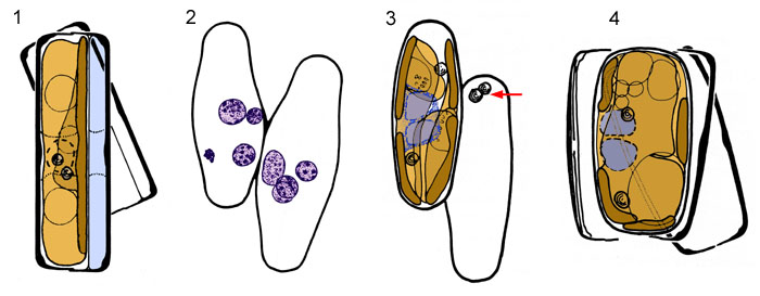
At meiosis I, each gametangium undergoes an unequal cytokinesis (Fig. 1), creating a larger cell beneath the hypotheca and a smaller, shallow cell beneath the epitheca (pale blue). The larger cell receives the whole of the chloroplast, both volutin granules and one of the meiosis I nuclei; the smaller cell receives only a nucleus (and its due proportion of the vacuoles of the parent cell).
Meiosis II takes place in both cells, so that four haploid nuclei are formed (Fig. 2). In the larger cell of each gametangium, one of the two haploid nuclei survives while the other contracts and degenerates; during this process it stains very intensely with nuclear stains (e.g. acetocarmine, haematoxylin), relative to the surviving nucleus, i.e. it becomes 'pyknotic' (bottom left in left-hand cell).
After meiosis II, the epitheca and hypotheca of each gametangium split apart and gape slightly in the zone where they abut, and an aperture is formed between them. This is shown on the next page. Fertilization occurs through fusion of the two large cells formed by the gametangia, and the movement of one of them out of its gametangium and into the other. The gametes, and hence the gametangia, can thus be regarded as 'male' and 'female', or 'active' and 'passive'. Because of this behavioural anisogamy, the zygote lies within the 'female' gametangium (Figs 3 and 4). The two volutin granules (red arrow, Fig. 3) of the male gamete and some residual cytoplasm are left behind in the male gametangium, which otherwise appears empty.
After plasmogamy, the male and female nuclei (dull purple in Figs 3 and 4) become closely associated on one side at the centre of the zygote, just beneath the zygote wall. They do not fuse until after the zygote has begun to expand.


 This site is hosted by the Royal Botanic
Garden Edinburgh.