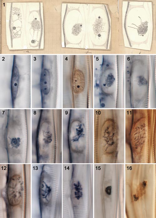
These images (apart from the drawings) were prepared in early 2013
from
Geitler's original slides. The microscope used was a Zeiss Axioimager,
with a 100× oil immersion objective (nominal N.A. 1.4) and interference
contrast optics. The variation in colour reflects differences in how
well the stain has lasted: around the edges of some preparations, the
stain has faded to pale brown or near transparency, whereas
nearer the centre, the original blue-black staining has often survived.
Cymbella neolanceolata:
meiosis

Figs 1–16. Meiosis in Cymbella neolanceolata, from material prepared in 1925 by Lothar Geitler. The material was obtained in summer 1925 from the Großer Plöner See, Germany, fixed with Schaudinn's solution, stained with Heidenhain's iron haematoxylin, and mounted in Canada Balsam.
Fig. 1. Drawings made by Lothar Geitler for his 1927 publication on meiosis and auxosporulation in what he called "Cymbella lanceolata" (left to right, the images were reproduced as Taf. 8, figs 2, 3 and 4).
Figs 2, 3. Nuclei of vegetative cells, containing prominent nucleoli.
Fig. 4. Leptotene (transition to meiosis).
Figs 5-8. Zygotene (synapsis).
Figs 9, 10. Entry to pachytene.
Figs 11, 12. Pachytene.
Fig. 13. Diplotene.
Fig. 14. Prometaphase.
Fig. 15. Metaphase of meiosis I.
Fig. 14. Telophase of meiosis I.
Click on the image for a larger version.
References:
Geitler, L. (1927). Die Reduktionsteilung und Copulation von Cymbella lanceolata. Archiv für Protistenkunde 58: 465–507.
Mann, D.G. (in press). Unconventional diatom collections. Nova Hedwigia, Beiheft.
David Mann,
Royal Botanic Garden Edinburgh
July 2013

 This site is
hosted
by
the Royal Botanic
Garden Edinburgh.