Recording images of diatoms
- Drawing
- Photomicroscopy
- Filters for black-and-white microscopy
- Digital microscopy
Drawing
back upIn the eighteenth and early nineteenth century, microscopists recorded what they saw of diatoms by drawing them freehand. With skill and practice, such drawings can be quite accurate, especially if measurements of the length and breadth of the diatom are made beforehand and used to constrain the drawing on squared paper or graph paper. However, just as few portraitists can make a good likeness of human beings, so very few microscopists can faithfully record the nuances of shape and pattern in diatoms - and we know from recent molecular genetic data and mating experiments that very subtle differences in shape can be highly significant [e.g. Behnke et al. 2004].
Nineteenth century drawings were reproduced for publication using etching, engraving, or lithography. Some examples are given on the DIADIST website.
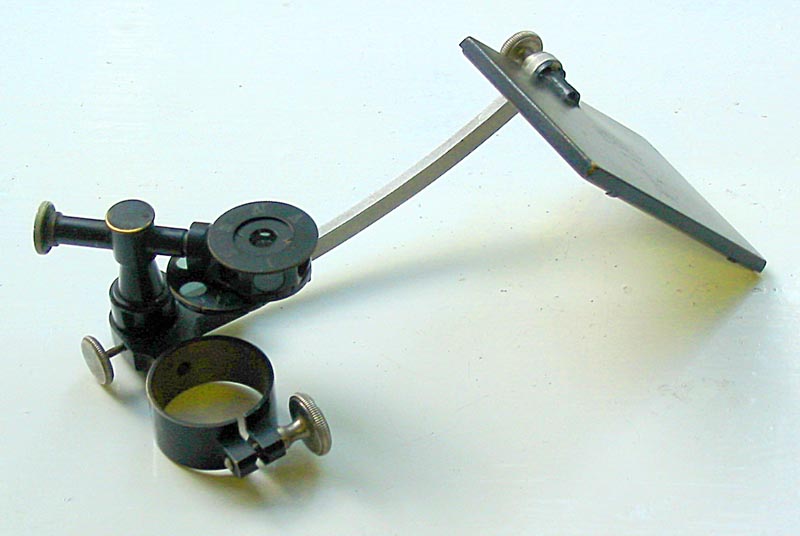
pre-WWII Carl Zeiss Jena camera lucida
Camera lucida
In the late nineteenth century various types of camera lucida drawing aids were introduced.
A common type consists of a prism that is placed over the eyepiece and a side-arm bearing a mirror, which allows the observer to simultaneously view the specimen and paper placed on the bench to the side of the microscope.
More sophisticated versions of the microscope camera lucida (such as the instrument illustrated), have filters both beneath the prism and in the light path between prism and mirror, to help balance the illumination between drawing and specimen.
The device fits around the tube of a monocular microscope and there is a prism in the vertically pivoted top arm which, when in place above the microscope eyepiece, allows simultaneous viewing of specimen and drawing: the prism comibines light from the microscope with light from the aligned, pivoted mirror on the side-arm. Two sets of graded filters are present, one in a ring below the prism (controlling the intensity of light from the microscope), the other in a cassette that rotates around the prism housing (controlling the intensity of light from the drawing). [click image for enlargement]
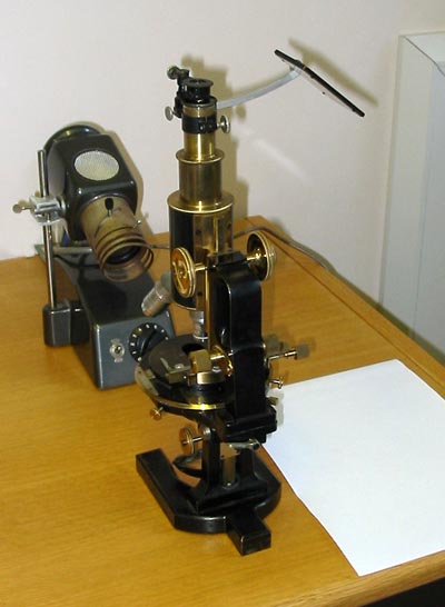
pre-WWII Carl Zeiss Jena microscope with camera lucida in place
I used this microscope for research published even since 1980! The quality of microscope lenses has not increased dramatically during the last 100 years. The main problem with instruments such as that shown is the physical discomfort of using them.
With suitable adjustments of the microscope illlumination and
the room lighting, it is possible to trace very accurately around the
virtual image of the specimen.
Almost all the drawings in my PhD dissertation (Mann 1978)
were done
using a Zeiss camera lucida, providing magnifications on the paper of
up to ×4350. The simple camera lucida must be used with care:
lateral distortion can occur if the angle of the mirror is incorrect
and there is evidence of this, for example, in some of the earlier
drawings in A. Schmidt’s Atlas der Diatomaceenkunde (see
example).
The camera lucida (referred to as a 'drawing attachment' by some modern
manufacturers) is not redundant. Drawing enables the observer to select
what is recorded, and it also makes it possible to combine information
from many different focal planes. In diatoms, this is especially useful
for recording details of sexual reproduction and auxospore development.